

Retinal Detachment
-
Retina Center
-
Retinal Detachment
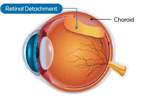
What is Retinal Detachment?
Retinal detachment is a condition in which the retina —
the neural tissue that should remain attached to the inner wall of the eye —
lifts and separates, typically due to vitreous traction.
If left untreated, it can lead to permanent vision loss
and is considered an ophthalmic emergency.
Normal Retina

Retinal Detachment

Normal Vision
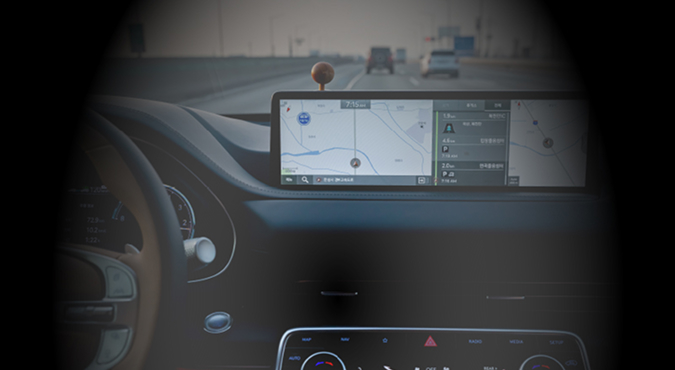
Vision with Retinal Detachment
Why Choose Our Retinal Detachment Surgery?
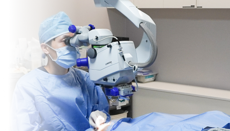
— A Skilled Retinal Specialist
The retina is an extremely delicate tissue, just 0.1-0.5mm thick,
located at the innermost depth of the eye.
Our retinal specialist, with 20 years of experience,
performs precise surgery that minimizes damage
to surrounding tissues.
Performed
From straightforward retinal detachments to recurrent
and highly complex cases, our record of over 10,000
successful surgeries is the cornerstone
of our clinic's ability to deliver consistently reliable outcomes.
Same-Day Registration ,
Same-Day Emergency Surgery
Retinal detachment is a race against time.
Surgery must be performed
before the detachment reaches the macula 'the center of vision'
in order to preserve sight.
Our clinic operates a fast-track system that allows
for immediate emergency surgery on the same day as the examination.
Air Tamponade Method Applied
Instead of conventional gas or silicone oil,
our clinic applies the air tamponade method in approximately 95% of all cases,
maximizing patient comfort and convenience.
Before Surgery
2 Days Post-Op
(Air 80%)
7 Days Post-Op
(Air 30%)
10 Days Post-Op
(Air 0%)
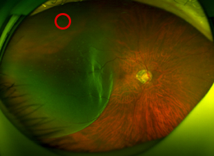
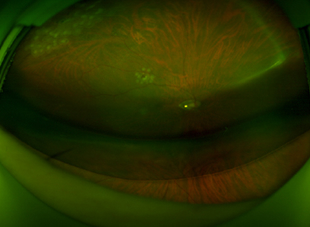
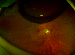
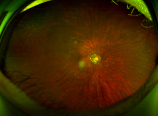
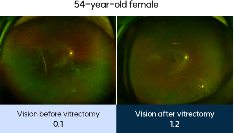
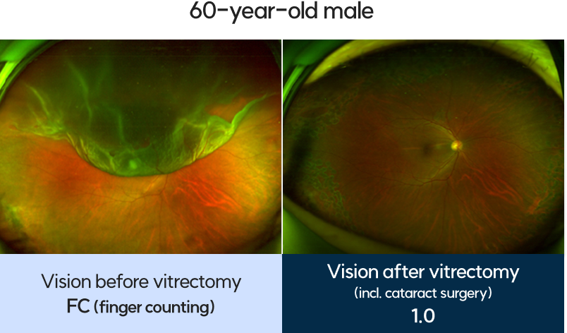
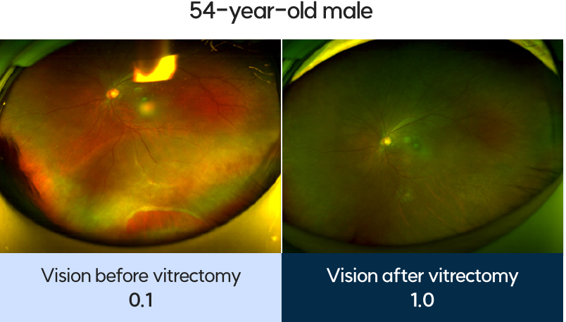
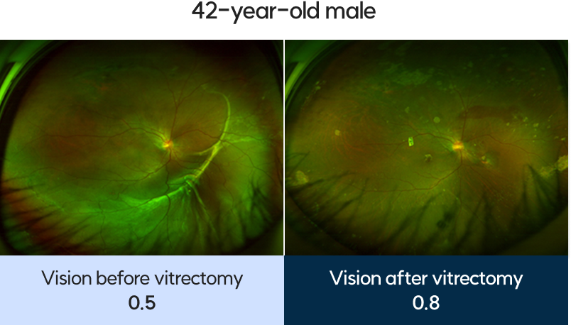
First Samsung Eye Clinic
Retinal Detachment Treatment Cases
Post-Operative Precautions
Proper post-operative positioning is just as important as a successful surgery.
the Correct Position
The buoyancy of the injected air is used
to press the retina back into place,
so patients must maintain a
face-down or side-lying position
for a specified period of time.
Process
The air is naturally absorbed
by the body at approximately 10% per day.
While air remains in the eye, vision may appear as
if looking through water —
this is a normal part of the recovery process.
Visits
To confirm that the retina
has reattached stably after surgery,
patients must visit our clinic according
the follow-up schedule provided.
With 20 years of expertise
and a record of 10,000 surgeries,
we are committed to protecting your
precious vision to the very end.
FIRST SAMSUNG EYE CENTER
